Hip Anatomy
The Anatomy of the Hip Joint
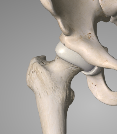
The hip is a bullet-like joint in the acetabulum. At the upper end of the femur is the spherical femoral head which is located inside the spherical acetabulum, which belongs to the pelvic bones. To limit friction between the surfaces and allow for easier movement, the opposite surfaces (articular surfaces) are covered with articular cartilage. The cartilage is lubricated by a small amount of yellowish fluid (synovial fluid) allowing the joint to move with even less friction than an ice skate on ice.
The femoral head is connected to the diaphysis of the femur, by a fixed bridge of bone called the femoral neck. When older people break their hips, it is the femoral neck that is destroyed. At the junction between the femoral neck and the diaphysis of the thigh there is a large bony bulge called the major trochanter. This is the hard bony protrusion that any person can feel when palpating the outside of their hip and which most patients usually show when they want to show pain in their hip. In fact, the major trochanter is not part of the hip joint, but is connected to the hip joint with the femoral neck and femoral head.

The femoral head is held inside the acetabulum by strong ligaments. If these ligaments are cut (for example in surgery) or torn (for example in a car accident) then the hip can be dislocated. The dislocation can destroy the blood supply to the femoral head by tearing the blood vessels, and the poor blood flow that comes as a result can in turn lead to arthritis in later years.
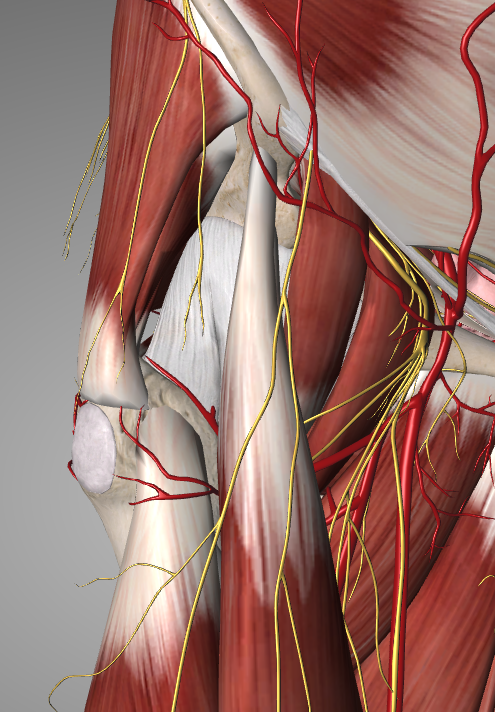
The hip joint is surrounded by three important nerves: the femoral nerve, the sciatic nerve, and the thyroid nerve. These nerves carry nerve impulses to and from the hip, groin and lower limbs to the brain through the spinal cord so that movements can be controlled and stimuli can be felt. These nerves in turn are surrounded by vessels and muscles. The most powerful muscles that support the hip joint are the three muscles known as the gluteus (major, middle and minor gluteus) behind and the rectus femoris and iliopsoitis in front of the hip joint. When the gluteal muscles weaken, as happens in advanced osteoarthritis, then the patient begins to limp.
The hip joint develops in the fetus as early as the first 8 weeks in the womb. Bones do not appear at first in the form of bone but start as cartilage (a stable gelatinous substance) and then gradually transform into bone (a hard substance). This process is known as ossification and involves the removal of cartilage, which is replaced by calcium-rich bone, which is produced by special osteoderived cells. The ossification centers of most bones ossify in childhood, but their limbs remain immature until puberty to allow growth. At some stage between the ages of 15 and 25 the hip joint is now fully developed and the increase in height in this area stops.
As people age, their bones may become thinner and in some cases smaller. This is known as osteoporosis, a form of bone weakening that can lead to fractures especially in the hip, wrist and spine. Osteoporosis is completely different from arthritis, yet sometimes these two entities are confused.

 Hip Osteoarthritis

 +30 210 7292002
 alilikakis@yahoo.com