Knee Anatomy
The Anatomy of the Knee Joint

The knee is a very complex joint which is formed by three bones: the tibia, the femur and the patella. The fibula is adjacent to the knee joint but is not part of it.
The knee is a hinge-like hinge, meaning it allows movement in a single plane for the most part, like a door hinge, although some degree of turning is also possible. The lower end of the femur is rounded and forms the femoral tubers. The upper end of the tibia is flattened and forms the tibial tubers. Logically, it is a miracle that a joint with this shape can have balance, but it achieves it with the support of the strong ligaments that connect the lower end of the femur to the upper end of the tibia. Knee ligaments include lateral ligaments, one on each side, and cruciate ligaments in the center of the joint. The cruciate ligaments, especially the anterior cruciate ligament, are the structures on which athletes rely most often and which they therefore injure most often.
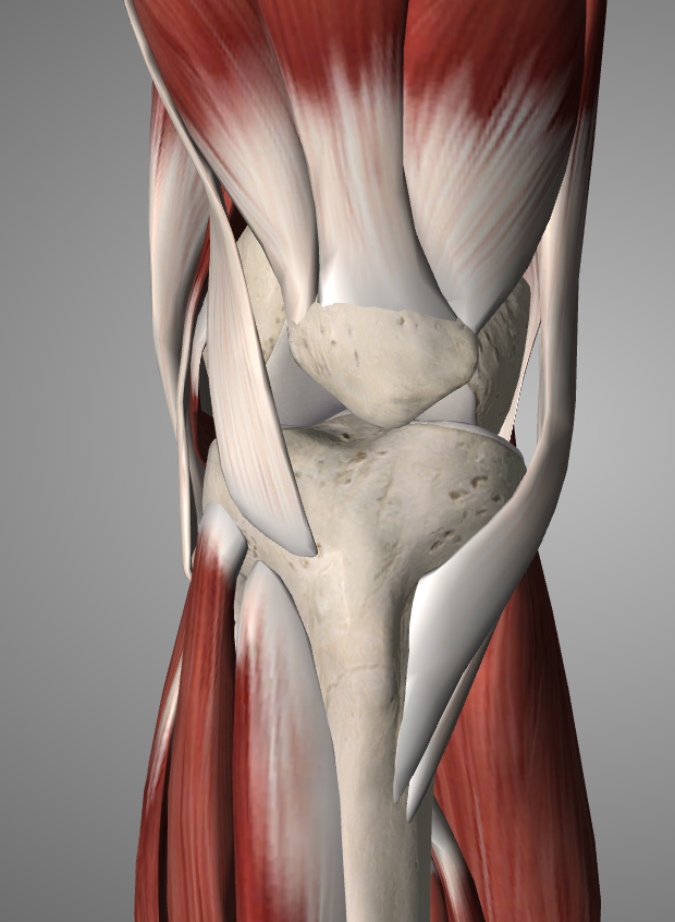
In the anterior part of the knee is the patella (“cup”). It is placed in front of the lower limb of the femur, forming with it a joint called the patellofemoral. Injuries and problems of this joint are a common cause of pain. At the upper end of the patella is the quadriceps (the large massive muscle in front of the thigh) and at its lower end, there is a tendon (patellar tendon) which is attached to the upper border of the tibia. The patella helps to strengthen the muscles responsible for the extension of the knee (“stretch”) which occurs when the quadriceps contracts and pulls the patella which in turn pulls, pulls the tibia. In essence, the patella is a fulcrum for the transfer of movement from the quadriceps to the tibia. Any activity involves extending the knee under pressure and by exerting force such as rapid ascent and descent of stairs with two steps by two, puts the kneecap under a heavy load.
As with the hip joint, the articular surface of the knee is covered by articular cartilage. And in the knee joint there is synovial fluid. There are many arteries around the knee that bring blood from the heart to the elements of the foot. Larger is the popliteal artery located just behind the joint and is a continuation of the femoral artery that has passed next to the hip a little higher.
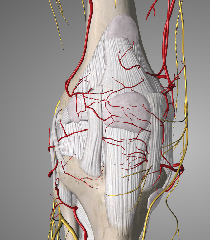
Two large nerves are also present in the knee joint. These are the tibial nerve behind and the common peroneal nerve on the outer side. These nerves and vessels are important because any trauma to the knee joint can also affect blood flow or nerve supply to the leg. The common peroneal nerve is particularly important because damage to it results in paresis of the foot (“drop” foot) in which the patient is unable to lift his fingers or foot upwards. This can often happen after a fracture, a problem in the spine (e.g. herniated disc), nerve pressure or as a complication of surgery.
As with the hip, the knee first presents itself as a cartilaginous structure and is later ossified. The cartilaginous femur can initially be seen in intrauterine life 8 weeks after conception while the tibia appears shortly after. The patella does not present in bone form at least until the age of 3 in girls and even later in boys. The knee joint as a whole stops growing at the ages between 17-20 years.
In the first years of life, the knees are usually softer and more flexible than later in life. They are harder to break and heal faster if they do. Sometimes childhood injuries can lead to arthritis in later years. There are also some types of arthritis that are only found in childhood, which in turn can lead to major surgeries at a young age.

 Hip Osteoarthritis

 +30 210 7292002
 alilikakis@yahoo.com