Arterial and Venous Thrombosis
PATHOPHYSIOLOGY OF ARTERIAL AND VENOUS THROMBOSIS

Injury to a blood vessel triggers a sequence of reactions: 1) vasoconstriction to reduce blood flow, 2) creation of a platelet embolus at the wound site that involves platelet attachment, activation and accumulation, and 3) blood clotting which includes a complex group of protease reactions. Platelets, nuclear cells with a lifespan of 7 – 10 days, come from the segmentation of megakaryocytes and are produced at a rate of 10 mg per day, a production that can be increased tenfold in cases of need. When the vessel wall is injured or the endothelium is disrupted, collagen of the subendothelial layer is exposed to the blood flow. Within seconds, platelets bind to the exposed collagen, which causes them to activate, degranulate and accumulate, which leads to the formation of a primary platelet plunge. At the same time, the coagulation mechanism is activated by the exposed hypoendothelial tissue factor (IP), resulting in the formation of fibrin, which stabilizes the platelet plunge. The pathogenesis of arterial thrombosis differs from that of venous thrombosis, a difference that is also reflected in the different strategies for dealing with them.
1a. VENOUS THROMBOSIS
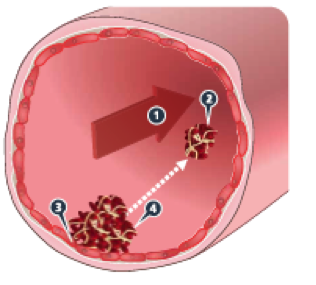
Venous thromboembolism (VTE) is the most common vascular disease after acute myocardial infarction (OEM) and stroke (stroke) and includes deep vein thrombosis (DVT) and pulmonary embolism (PE, PE). DVT most often occurs in the large veins of the lower extremities. When part of the clot is detached, it can migrate to the lungs and disrupt or cut off blood flow to the pulmonary artery. More than 150 years ago, Rudolph Virchow proposed a triad of causes of venous thrombosis: venous stasis, injury to the vascular endothelium, and hypercoagulability of the blood. This trio is still applied with one or more actors involved in the development of DVT.
Venous stasis (or reduced blood flow to the veins) can be caused by many causes, including immobility (e.g., hospitalization or nursing home), major orthopaedic procedures (e.g., during and/or after surgery), or increased venous pressure (e.g., heart failure). The majority of venous clots form in areas with slow blood flow, and reducing venous posture in the legs has been found to reduce the risk of VTE. Venous posture promotes clot formation by failing to quickly remove activated clotting factors from the site of vascular injury. In addition, local hypoxia and vascular wall dilatation can activate the endothelium, causing the expression of the selectin adhesion molecule on its surface. Newer data show that microparticles – carriers of the tissue factor may play an important role in venous thrombosis, similar to platelets in arterial thrombosis. Factors that may allow a small, silent clot to grow, or a new clot to grow, include prolonged disruption of venous function, maintenance of hypercoagulability, and damage to the endogenous anticoagulant or fibrodolytic system. Venous clots consist mainly of red blood cells and large amounts of fibre. The activation of the coagulation system is the primary cause of venous thrombosis and precedes the activation and aggregation of platelets. This explains why anticoagulant therapy is the primary strategy for dealing with VTE and the guidelines emphatically recommend the use of anticoagulants for the prevention and treatment of VTE.
VTE is a common and significant complication of atrial fibrillation (AF) and as a result these patients take preventive anticoagulants, mainly to avoid stroke. Virchow’s triad is also applied to the formation of clots in the AF with the venous position (especially in the vagina) being the most determining factor. AF also inhibits the contractile capacity of the vagina through multiple mechanisms, and the restoration of contractility leads to thromboembolism. There is also evidence of dysfunction of the endothelium of the vagina in AF, while biomarkers indicate a prothrombotic role of local inflammation at the same time as changes in the coagulation system.
1b.ARTERIAL THROMBOSIS
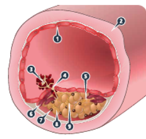
Triggering arterial thrombosis is the rupture of an atherosclerotic plaque that causes complete or partial blockage of the vessel, usually in tissues with terminal vascularization. Indeed, cardiac ischemia and stroke are the most serious clinical manifestations of arterial thrombosis. When atherosclerotic plaque rupture occurs, its fatty nucleus is exposed to the blood circulating in the arterial lumen. The core area of the plaque contains tissue factor and collagen pieces, which are highly thrombogenic. Circulating BP also increases in patients with cardiovascular disease and may contribute to thrombosis after a plate rupture. At the initial stage of rupture, platelets rapidly accumulate in the area, followed by further aggregation and rapid growth of the clot. The coagulation cascade is activated at this stage by the formation of thrombin, which also activates platelets. Activated platelets promote their further aggregation, adhesion, aggregation and activation. Therefore an arterial clot is rich in platelets and exposed to rapid blood flow. The fibrous network of the clot increases as it expands within the arterial lumen, and its surface is covered by activated platelets. In the last stage of thrombosis, a loose fibral network with large numbers of trapped red blood cells complete the formation of the clot.
Although antiplatelet agents are commonly used for the prevention and treatment of arterial thrombosis, there is a clear activation of the coagulation mechanism after the rupture of the atherosclerotic plaque, which provides a mechanistic rationale for anticoagulant therapy. Clinical evidence suggests that, in some cases, the combination of anticoagulant and antiplatelet therapy is more effective than either treatment on its own.
- RISK FACTORS FOR ARTERIAL AND VENOUS THROMBOSIS
The classic risk factors for arterial thrombosis are presented in Table I and for venous thrombosis in Table II.
TABLE I: Risk factors for arterial thrombosis.
| Hyperlipidemia | 3,25 (2,81-3,76) | Smoking | 2,87 (2,58-3,19) | Diabetes | 2,37 (2,07-2,71) | Hypertension | 1,91 (1,74-2,10) | Abdominal obesity | 1,62 (1,45-1,80) |
TABLE II: Risk factors for venous thromboembolism
High-risk factors
|
Moderate risk factors
|
Weak risk factors
|
There is an exponential increase in the risk, both of arterial and venous thrombosis, with advancing age. Hereditary deficiencies of antithrombin and proteins C and S are rare but important risk factors for venous, but not arterial thrombosis, causing a significant reduction in the natural anticoagulant system and promoting thrombin production, thus causing susceptibility to VTE. The two most common genetic risk factors for VTE are mutations in the factor V and prothrombin genes, which also increase the risk of arterial thrombosis but to a lesser extent. Antiphospholipid antibody syndrome is one of the most important risk factors for both arterial and venous thrombosis. Similarly, a common risk factor, but mild, is hyperhomocysteinemia, which is due to genetic and acquired factors. There is increasing evidence for a correlation between metabolic syndrome and arterial thrombosis, but also venous thrombosis, to a lesser extent [metabolic syndrome is defined as the presence of at least three of the following diagnostic criteria: abdominal obesity, triglycerides, ̄HDL, AP, ED]. A previous episode of DVT is an independent and the most important risk factor for recurrence of DVT. Trauma, surgeries, and immobility are associated with an increased risk of thromboembolic disease, most likely through a mechanism of venous stasis and BP accumulation. Arterial thrombosis can be a manifestation of iatrogenic surgical injury or the first manifestation of immune thrombocytopenia (HIT II). Cancer is one of the most important acquired risk factors for VTE. Oral contraceptives increase the relative risk of venous thrombosis two to six times, as well as arterial thrombosis, causing endothelial dysfunction.
Various models for scoring venous thromboembolism risk factors in surgical patients have been presented in the literature. Caprini’s grading is characteristically mentioned, one of the oldest and most used, not validated for orthopedics anyway. The correlation of the risk score with the proven incidence of VTE in surgical patients is well documented.

 Hip Osteoarthritis

 +30 210 7292002
 alilikakis@yahoo.com