Rupture of labial cartilage
What is defined as a ruptured labial cartilage?
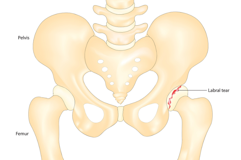
In the hip joint there is a fibrocartilaginous ring that surrounds most of the circumference of the acetabulum, which is called the labial cartilage. There are several similarities of the labia cartilage of the hip with the menisci in the knee, similarities both anatomical and functional. The rupture of the labial cartilage is an injury to this fibrocartilaginous ring.
The labial cartilage acts as a protective lining of the acetabulum. More specifically, it contributes decisively to the stability of the joint, increasing its surface area and maintaining a watertight environment between the acetabulum and the femoral head. In this way, direct contact of the bone surfaces is prevented, ensuring smooth hip mobility.
Additionally, labial cartilage reduces friction between bones, protecting them from premature decay.
When the labial cartilage ruptures, the normal mechanics of the joint are significantly disrupted and symptoms occur. Early diagnosis and treatment are crucial to prevent further damage and maintain joint health.
What are the causes of its cause?
A very common cause of rupture is the overuse of the joint, either due to sports activities or due to demanding movements in everyday life or work. In addition, a hip injury in a sport, fall or accident can cause an acute rupture.
In several cases, the lesion arises as a result of underlying anatomical abnormalities, such as femorocoililiary impingement syndrome or hip dysplasia. Finally, joint degeneration due to hip osteoarthritis can also lead to rupture of the labial cartilage. It is noteworthy that the relationship between a ruptured labial cartilage and osteoarthritis is bidirectional: one condition can cause or aggravate the other.
Which patients are more likely to have a rupture?
Rupture of the labial cartilage of the hip most often occurs in people who repeatedly strain the hip joint. In particular, this strain is mainly caused by sports or professional activities. Those involved in activities involving sharp rotational movements or large-scale trajectories, such as martial arts, football, golf and dance, especially classical ballet, are at increased risk.
Intense strain and repetitive micro-injuries can cause gradual wear and tear of the labial cartilage.
With what symptoms does it appear?
In several cases , the rupture of the labial cartilage may remain asymptomatic.
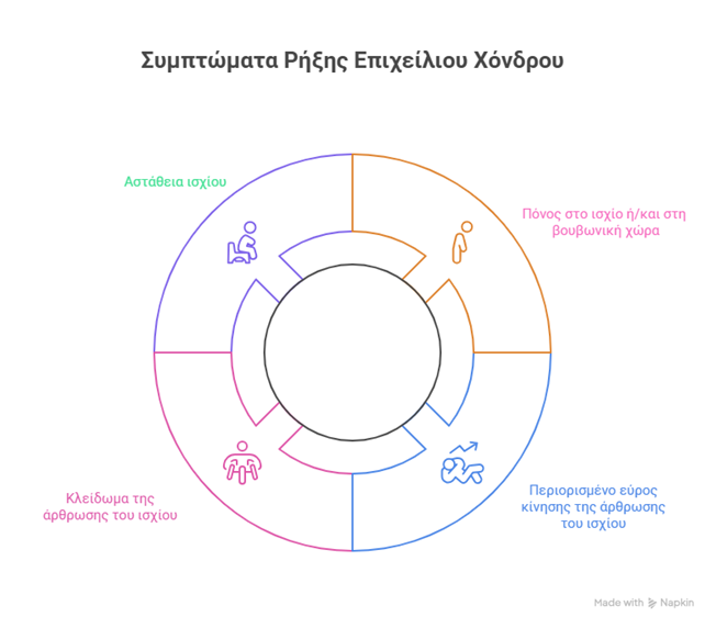
However, when clinical manifestations occur, the most common symptoms include:
- Pain in the hip and/or groin area. This pain is often aggravated by walking, standing for long hours or prolonged immobility.
- Limitation of the range of motion of the joint, with difficulty in movements such as flexion or medial rotation of the hip.
- Feeling of “locking” the joint during movement.
- Feeling of instability in the hip, especially in more advanced stages.
How is the diagnosis made?
The diagnosis of a labial cartilage rupture is initially based on taking a detailed medical history and a detailed clinical examination by a specialized orthopedist. During the examination, the mobility of the hip joint is evaluated through special tests that may reproduce the symptoms.
However, confirming the diagnosis requires imaging testing. Simple X-rays can reveal any concomitant lesions. On the other hand, MRI, especially when accompanied by intra-articular contrast injection (MRI arthrogram), allows the imaging of soft tissues and labial cartilage more accurately.
However, the complex anatomy of the hip makes diagnostic imaging sometimes unclear. In doubtful cases, an intra-articular anesthetic can be administered, and if the symptoms subside, damage to the labrum cartilage is considered the most likely cause of the pain.
What is the appropriate treatment for ruptured labial cartilage?
Conservative treatment is mainly applied to patients with mild symptoms. It is also applied in cases where degenerative lesions, such as osteoarthritis or anatomical variations, such as hip dysplasia, coexist.
The aim of this approach is to reduce symptoms and improve hip functionality. The treatment plan includes:
- Rest
- Modification of aggravating activities
- Medication with analgesics and non-steroidal anti-inflammatory drugs
- Intra-articular injections of corticosteroids or platelet-enriched plasma (PRP)
At the same time, physiotherapy plays a crucial role, helping to strengthen muscles and improve hip range of motion.
In cases where the symptomatology is intense, persists despite the conservative approach or significantly affects the patient’s functionality, surgical intervention with the method of hip arthroscopy becomes necessary.
It is a minimally invasive technique, which allows access through small incisions. Damage repair is performed using specialized arthroscopic instruments and a high-definition intra-articular camera, which provides direct and detailed visual imaging of the joint.
The choice of the appropriate surgical strategy (suturing or partial resection and rejuvenation of the labial cartilage) depends on the type, location and extent of the rupture, but also on the age and requirements of the patient.
The operation has a high success rate in reducing symptoms and functional recovery of the hip. The patient is usually discharged from the hospital on the same day and follows an individualized rehabilitation program, with the aim of safely and gradually returning to daily activities.
If you experience symptoms that indicate a ruptured labial cartilage in the hip, trust a physician with years of experience and leadership in the field of reconstructive surgery. Orthopaedic Surgeon Anastasios Lilikakis is the Director of the Third Orthopaedic Clinic of the Athens Euroclinic and the President of the Department of Hip & Knee Reconstructive Surgery of EEHOT. Contact us and book your appointment to receive personalized treatment.
 Hip Osteoarthritis

 +30 210 7292002
 alilikakis@yahoo.com